Deep Vein Thrombosis & Pulmonary Embolism Treatment in India 2026
Deep Vein Thrombosis & Pulmonary Embolism Treatment in India 2026
Deep vein thrombosis (DVT) and pulmonary embolism (PE) — collectively known as venous thromboembolism (VTE) — represent one of the most underdiagnosed and underestimated cardiovascular emergencies across sub-Saharan Africa and North Africa. A clot forming in the deep veins of the leg can travel to the lungs within hours, causing a life-threatening pulmonary embolism. Yet the interventional tools to dissolve large clots rapidly, restore blood flow, and prevent catastrophic outcomes are available in India at a fraction of Western prices.
This guide covers everything African patients need to know about DVT and pulmonary embolism — diagnosis, treatment options, costs, and how to access India's vascular interventional radiology capabilities.
Key figures: Catheter-directed thrombolysis in India costs $4,000–8,000 versus $25,000–60,000 in the USA. IVC filter placement costs $2,500–5,000 in India. India's vascular interventional radiology centres manage DVT and PE using the same protocols as leading Western centres.
Understanding DVT and Pulmonary Embolism
What Is Deep Vein Thrombosis?
DVT occurs when a blood clot (thrombus) forms in one of the deep veins, most commonly in the leg (femoral or popliteal vein) or pelvis (iliac vein). Symptoms include:
- Swelling of one leg (unilateral leg swelling is the most characteristic sign)
- Pain or tenderness in the calf or thigh
- Redness and warmth over the affected vein
- The leg may feel heavy
DVT can develop without symptoms (silent DVT), which is why it is frequently missed.
What Is Pulmonary Embolism?
If a DVT clot breaks off and travels through the bloodstream to the lungs, it becomes a pulmonary embolism. The clot blocks a pulmonary artery, reducing oxygen delivery to the lung tissue. Symptoms include:
- Sudden shortness of breath (the most common symptom)
- Sharp chest pain, especially with breathing (pleuritic chest pain)
- Coughing, sometimes with blood (haemoptysis)
- Rapid heart rate (tachycardia)
- Dizziness or loss of consciousness (in massive PE)
Massive PE — where a large clot obstructs the main pulmonary artery — is immediately life-threatening and requires emergency intervention.
DVT and PE in Africa: The Rising Burden
DVT and PE are rising across Africa for multiple reasons:
Increasing risk factors: The epidemiological transition in Africa has brought rising rates of obesity, type 2 diabetes, hypertension, and metabolic syndrome — all of which increase clotting risk.
Surgery and hospitalisation: Major surgery is a potent DVT trigger. As more Africans undergo major operations, post-operative DVT risk rises.
Long-haul air travel: The boom in African intercontinental travel — particularly the growing medical travel corridor to India — increases exposure to immobility-associated DVT risk. This is sometimes called "economy class syndrome."
Sickle cell disease: Common in sub-Saharan Africa, sickle cell disease is associated with significantly increased risk of VTE.
Underdiagnosis: Many DVT cases in Africa go undetected because Doppler ultrasound of the leg veins — the standard diagnostic test — is not widely available at all hospitals and clinics.
Limited interventional options: Most African hospitals can manage DVT with anticoagulant drugs, but cannot offer catheter-directed thrombolysis, IVC filter placement, or other interventional vascular procedures.
Diagnostic Approach to DVT and PE
Diagnosing DVT
The gold standard investigation is compression Doppler ultrasound of the leg veins — a non-invasive ultrasound examination where pressure is applied over the vein. A normal vein compresses completely; a vein containing a clot does not. This test is accurate, rapid, and widely available at Indian hospitals (cost: $50–120).
For suspected pelvic DVT (iliac vein thrombosis), CT venography or MRI venography may be needed.
Diagnosing Pulmonary Embolism
CT pulmonary angiography (CTPA) is the definitive diagnostic investigation — a CT scan of the chest with intravenous contrast that images the pulmonary arteries directly and shows clots as filling defects. CTPA cost in India: $150–300.
For risk stratification, the treating team will also perform echocardiography to assess right heart function (right ventricular strain indicates more severe PE) and measure troponin and BNP blood markers.
Treatment Options for DVT and PE in India
Anticoagulation (Blood Thinners)
Anticoagulation is the cornerstone of DVT and PE treatment. It does not dissolve the existing clot but prevents it from growing and allows the body's natural fibrinolytic system to slowly break it down.
Options include:
- Low molecular weight heparin (LMWH): Injected subcutaneously once or twice daily. Used acutely during hospital admission.
- Direct oral anticoagulants (DOACs): Rivaroxaban, apixaban, or edoxaban — taken as tablets, no monitoring needed, highly effective. These have largely replaced warfarin for most patients with VTE.
- Warfarin: Older oral anticoagulant requiring regular INR blood test monitoring. Still used in certain patient groups.
For uncomplicated DVT or non-massive PE, anticoagulation alone is the standard treatment. Most patients can be managed as outpatients or with a short hospitalisation. Anticoagulation costs in India: $200–600 per month depending on the agent.
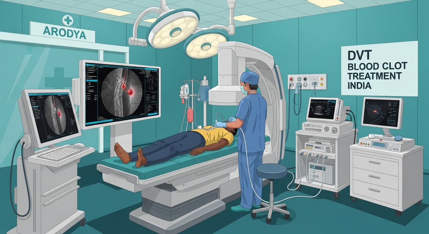
Catheter-Directed Thrombolysis (CDT)
For large proximal DVT (clot in the femoral or iliac vein extending into the pelvis) or submassive/massive PE, catheter-directed thrombolysis offers a faster and more targeted approach to clot removal.
An interventional radiologist inserts a fine catheter through a vein in the groin or neck and advances it directly into the clot under fluoroscopic guidance. A thrombolytic drug (alteplase) is then infused through the catheter directly at the clot site over twelve to twenty-four hours. This dissolves the clot far faster than anticoagulation alone, restoring venous blood flow and reducing the risk of post-thrombotic syndrome (long-term leg swelling and pain that affects 20–50% of DVT patients).
Who benefits most from CDT:
- Patients with iliofemoral DVT (large clots in the iliac or femoral vein) causing severe leg symptoms
- Patients with submassive PE (moderate right heart strain on echo) who are not improving on anticoagulation alone
- Young, fit patients at low bleeding risk where preserving venous valve function is important for long-term leg health
Cost of CDT in India: $4,000–8,000 including catheter, thrombolytic drugs, fluoroscopy suite time, monitoring, and two to three days ICU-level observation.
IVC Filter Placement
An inferior vena cava (IVC) filter is a small mesh cage inserted into the IVC (the main vein returning blood from the legs to the heart) that catches any clot travelling upward from the legs before it can reach the lungs. It is used when:
- Anticoagulation is contraindicated (recent surgery, active bleeding, brain haemorrhage)
- Despite therapeutic anticoagulation, PE recurs
- As a temporary measure in very high-risk surgical patients
Modern retrievable IVC filters can be removed once the DVT risk has resolved (typically after three to six months), avoiding the long-term complications of permanent filters.
Cost of IVC filter in India: $2,500–5,000 including the device, insertion procedure, and fluoroscopy.
May-Thurner Syndrome
May-Thurner syndrome (also called iliac vein compression syndrome) is a specific anatomical cause of left-sided DVT, where the right iliac artery compresses the left iliac vein. This obstruction causes venous stasis and predisposes to clot formation. Treatment involves catheter-directed thrombolysis followed by balloon venoplasty and stent placement in the compressed vein.
This condition is underrecognised in Africa and often explains recurrent left-leg DVT in younger patients. India's vascular interventional radiology teams are experienced in diagnosing and treating May-Thurner syndrome. Procedure cost: $5,000–9,000.
Long-Term Anticoagulation Planning
After DVT or PE treatment in India, you will return home on long-term anticoagulation. Planning for this is important:
DOAC availability: Rivaroxaban and apixaban are widely available in most African countries (though cost varies). Your Indian doctor will prescribe a regimen that is accessible where you live.
Monitoring: DOACs do not require routine blood monitoring — a significant advantage for patients returning to areas with limited laboratory access. Warfarin requires regular INR testing (every two to four weeks when stable), which your local clinic can manage.
Duration: Provoked DVT (triggered by surgery, travel, or temporary risk factor) typically requires three to six months of anticoagulation. Unprovoked DVT or PE — or recurrent events — may require twelve months or indefinite treatment. Your Indian vascular team will specify the duration in your discharge letter.
Thrombophilia testing: If you are young, had an unprovoked clot, have a family history of clotting, or experienced recurrent events, testing for inherited thrombophilia (such as Factor V Leiden, prothrombin gene mutation, antiphospholipid syndrome, or protein C/S deficiency) is recommended. This can be done in India before or after treatment.
DVT Prevention After Medical Travel to India
The long-haul flight to India (typically 8–12 hours from East Africa, 10–14 hours from West Africa) itself carries DVT risk, particularly for patients who already have vascular disease or are recovering from surgery. Protective measures include:
- Graduated compression stockings (below-knee, class II) worn during the flight
- Rising and walking the aisle every two hours
- Staying hydrated — avoid alcohol and excessive caffeine
- Ankle pumping exercises (circling the feet, flexing and extending the ankles) in your seat
- For very high-risk passengers (previous DVT, recent surgery, known thrombophilia), your doctor may recommend a prophylactic dose of LMWH before the flight
Arodya advises all patients on DVT prevention as part of our pre-travel briefing.
To discuss your DVT or PE situation with our coordination team and receive a hospital referral matched to your clinical needs, start your enquiry with Arodya. India's vascular interventional radiology teams can provide options that are unavailable or unaffordable in most African healthcare settings.
For information on how to plan your trip financially, see our budget medical trip guide.